;%20?%3E)
×

;%20?%3E)
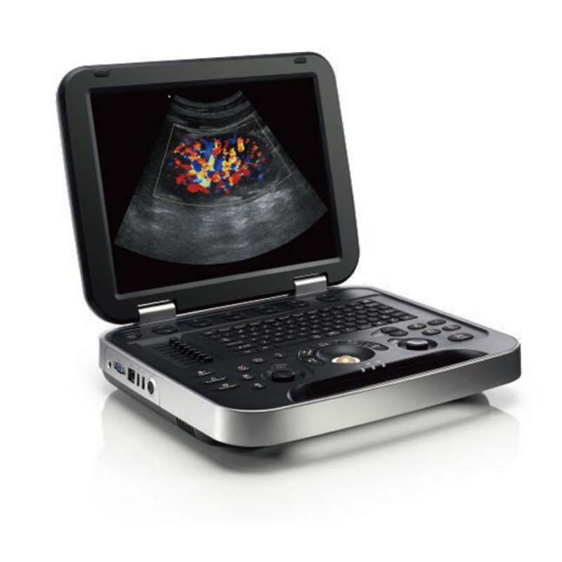
Excellente performance pour vous
En combinant un contrôle précis et adaptatif avancé de la transmission et de la réception avec une technologie de traitement parallèle multicanal efficace, la vitesse de calcul de la machine est maximisée tout en obtenant les meilleures performances d'image.
comme la réflexion spéculaire, le speckle, la diffusion et l'atténuation, assurant une image de qualité supérieure
clarity.
| PARAMÈTRE | CARACTÉRISTIQUES |
| Application: Générique, Abdomen, Obstétrique, Gynécologie, Vasculaire, Petites pièces, Urologie,
MSK , Cardiologie. |
|
| Types de transducteurs | Réseau convexe, réseau linéaire, micro-convexe, sonde intracavité. |
| Modes de fonctionnement | Mode B, Double B, Quadruple B, Mode CF, Mode M, Mode B/M, Mode PW
Imagerie Doppler de puissance, imagerie Doppler de puissance directionnelle Vue large (Étendre le champ de vision) (Optionnel), Image dans l'image Trapèze, Panorama, Mode couleur M, Mode anatomie M Mode d'amélioration de l'aiguille (optionnel), mode d'amélioration du contraste (optionnel) |
| Techniques et fonctions d'imagerie | Optimisation automatique
Imagerie spatiale composée Imagerie par réduction du speckle Quantification des couleurs direction à angle fin Imagerie harmonique tissulaire (THI), imagerie harmonique par inversion d'impulsion (iTHI) convexe virtuel Base de données d'informations sur les patients Archive d'images sur disque dur intégré Calcul Doppler automatique en temps réel calculs OB Tendances fœtales Calculs multi-gestationnels calculs de dysplasie de la hanche Calculs gynécologiques calculs vasculaires calculs urologiques Calculs rénaux Documentation électronique embarquée PW Auto-trace Confidentialité et sécurité DICOM Vue panoramique Auto IMT connexion imprimante USB externe |
| Capacité simultanée | B/CF, B/PDI ou DPDI, B/PW, C/M, B/M, B+CF ou PDI ou DPDI/PW ou M. |
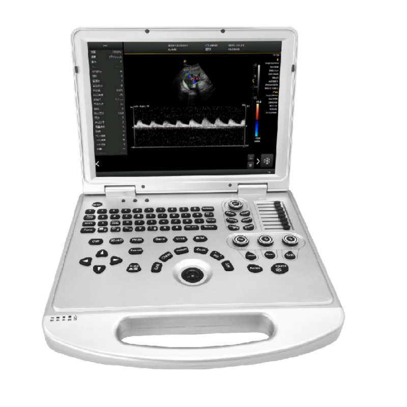
| Écran tactile | Moniteur LED haute résolution de 15 pouces
Résolution: 1024 * 768 bouton en silicone design Fermeture magnétique Haut-parleur intégré Réglage de la luminosité et du contraste |
| Sonde convexe | Applications :
Médecine générale, abdomen, obstétrique, gynécologie, urologie |
| fréquences:
2.5, 3.5, 4.0, 5.0, 6.0, H3.6, H4.0, H5.0, H6.0 MHz |
|
| Sonde linéaire | Applications :
Petites parties, vasculaires, musculo-squelettiques, superficielles |
| fréquences:
5.0, 7.5, 8.5, 10.0, H8.0, H9.0, H10.0, H12.0 MHz |
|
| Sonde transvaginale | Applications :
Obstétrique, gynécologie, urologie |
| fréquences:
4.0, 5.0, 6.5, 8.0, 9.0, H7.0, H8.0, H9.0 MHz |
|
| Sonde microconvexe | Applications :
Médecine générale, abdomen, obstétrique, gynécologie, urologie |
| fréquences:
2.5,3.5,4.0,5.0,6.0,H3.6,H4.0,H5.0, H6.0 MHz |
|
| Sonde transrectale | Applications :
Obstétrique, gynécologie, urologie |
| fréquences:
5.0, 7.5, 8.5, 10.0,H8.0,H9.0, H10.0, H12.0 MHz |
|
1. Configuration standard :
Câble d'alimentation, une sonde convexe, manuel d'utilisation.