;%20?%3E)
×


;%20?%3E)
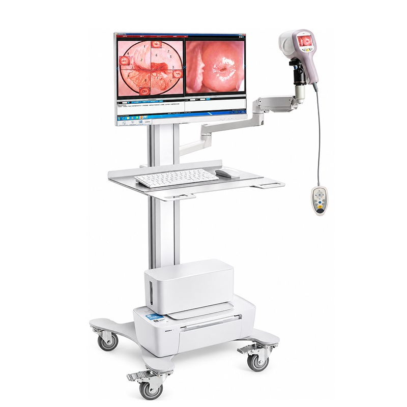
Cela consiste en colposcope camera, external capture card, trolley with integrated arm , software with key, foot switch, control handle.
-Latest full HD technology colposcope use 1/2.8 inch Sony CCD, 3.27 million effective pixels, 30 times ,optical zoom, with high-quality amplification effect can be clearly observed a variety of small pathological cells, can be accurately positioning the biopsy of the lesion site, help improve the diagnosis of various cervical diseases, pre-cancerous screening accuracy, reduce missed Diagnosis.
-Advanced spot subtraction imaging technology and optical image stabilization technology, reduce the light spot caused by the phenomenon of reflection and blurred images by lens’ shaking and offsetting. Ø
-5” LCD screen, Real-time dynamic images can be observed. Combined with built-in battery, used in rapid inspections and large-scale census work, make examination more quick and convenient.
-Fully-damped camera holder, 360° full-range adjustment, automatic hovering at any angle
-All-metal material, integrated design, more convenient to move, suitable for all types of inspection room.
-Flexible positioning of the swing arm bracket. The position and angle of the lens can be adjusted arbitrarily, to meet the needs of clinical examination and operation, to provide doctors with no dead angle of observation vision.
-Four imaging modes highlight important diseased tissues and blood vessels, epithelial tissues, and truly restore tissue primary colors and levels of detail to meet different clinical needs.
| PARAMÈTRE | DONNÉES TECHNIQUES |
| CAMERA SET | |
| Capteur d'image | 1/2.8 inch Sony CCD |
| Efficace à partir de pixels | 3.27 millions de pixels |
| Taille de l'image | 1920*1080 |
| Résolution horizontale | 1080P/25; 1080P/60 |
| Image output mode | 3G-SDI, Transmission rate: 2.97Gb/S |
| Temps de grossissement | System magnification time: 240 times |
| Optical magnification: 20 times | |
| Digital magnification: 12 times | |
| focus mode | Fast autofocus, Manual focus, Manual focus support independent key control macro adjustment function |
| Profondeur de champ | 4.3mm ~ 129mm |
| Distance de travail | 200 ~ 400mm |
| Champ de vision | Ф16 ~ 480 mm |
| Saturation de couleur | 95 % |
| Image geometric distortion | ≤2.5% |
| Light soruce | Energy-saving and environmentally protection 20 groups of white LED light sources, high brightness, long life, Illuminance uniformity, high color rendering index, with no heat and so on. |
| Lifetime of light source | heures ≥50000 |
| Illuminance of light source | ≥1200 XNUMXLux |
| Gamme d'illumination | Ф≥80mm |
| Color temperature of light
source |
5000K ~ 5500K |
| Indice de rendu des couleurs | ≥95, restore the true tissue color and level of detail |
| Augmentation de la température de surface | ≤41 ° C |
| Mode d'imagerie | Colorless imaging: highlights important lesions and blood vessels, helping doctors quickly retrieve diseased tissue. |
| Natural light imaging: low color temperature imaging technology, softer light, help doctors better observe epithelial tissue | |
| LED light imaging: high-definition LEDs with unique imaging technology to restore the original color and level of detail | |
| Filter mode: The four-level green mode enhances the recognition of tiny blood vessels and highlights blood vessel details. | |
| Technologie d'imagerie | Dark light imaging technology: Color images can be clearly observed in dimly lit examination room. |
| Light spot reduction technology: In the observation of tissues with more body fluids, can reduce the light spot caused by the reflective phenomenon | |
| Optical image stabilization technology, reduce blurred images by lens’ shaking and offsetting. | |
| Status mode display | Current magnification, focus mode, freeze mode, imaging mode, green mode status display, and acetic acid reaction and iodine staining test time display, controlled by the handle button |
| Poignée de commande | The palm handle design, user-friendly operation panel, silicone button oval layout, zoom, focus, imaging mode, acetic acid timing, image marking, brightness adjustment and other functions can be completed with one hand. |
| Alimentation | Power adapter: DC12V |
| Internal battery: Internal battery power supply mode, used with back 2.5’’ screen
Specifications: 7.2V 2.1Ah; charging time: ≥ 4 hours; battery life: 3 hours(without screen), 2.5 hours (with screen) |
|
| Optionnel
|
USB capture device :used with the notebook, easy to carry and convenient for gynecological census work |
| Video converter box:connect the monitor directly,apply to doctorsinstructional video. | |
| Bar code gun: It is convenient for doctors to quickly find case information and quick review, and better realize hospital information management | |
| Computer with monitor | |
| Imprimante | |
| CONFIGURATION STANDARD | |
| Caméra | 1/2.8 inch Sony CCD;3.27 million pixel |
| Support de caméra | Full-dampness camera holder, 360-degree adjustment, automatic stop at any angle, with damping fine-tuning function |
| Interrupteur au pied | IPX1 waterproof foot switch with image capture and video control |
| Manche télescopique | Single screen :1062*818*1368mm (including screen) |
A multi-functional management system integrating image acquisition, image processing, clinical diagnosis, report editing, and query statistics. U disk automatic installation, multi-user operation mode, easy to use for different doctors. Support WIN7 operating systems above with Chinese, English, Spanish, Russian, Vietnamese, Turkish etc.
1) Unique image capture function:
With foot switch and software two ways of capturing, recording, and can realize dynamic, static, full screen image capture, 100 color images can be collected at each check.
2) Powerful image processing function:
With dynamic and static image comparison function, can be compared with standard image, and the collected images can be zoomed in, zoomed out, negatively imaged, marked, text annotated, partial magnified , area perimeter calculated etc., which is more convenient for doctors to identify the diseased tissue
3) Professional clinical diagnostic function:
Professional case database (up to 175), satisfied cases images can be added, suitable
for clinical contrast. The professional clinical diagnosis knowledge database provides
a large number of editable clinical terms. With RCI automatic assessment system to
make the inspection more objective and accurate.
4) Rich report editing function:
Built-in 19 standard case report template, convenient for editing and printing. It can
display 1-6 pictures in the same screen. The report template can be designed, modified, and output in PDF format. Provides evidence for prevention of medical disputes.
5) Convenient and quick query statistics function:
Search cases via case number, name, bar code, date of birth and date of examination;
Image display, data modification, save, re-examination and video reply to the retrieved cases. Convenient for hospital information management, improve management efficiency.


